


Dunedin Brain Imaging Study MRI Protocol
Hippocampus Paradigm
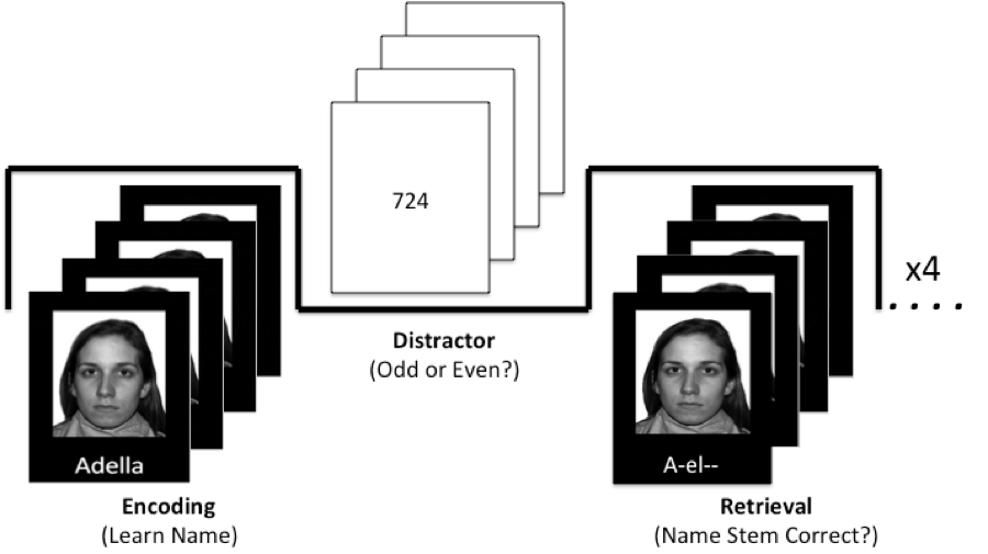
Hippocampus Reactivity Paradigm (Face-name Task)
Our fMRI paradigm consists of the encoding and subsequent recall of novel face-name pairs (Zeineh et al., 2003). A distractor task (odd/even number identification) is interleaved between encoding and recall blocks to prevent maintenance of information in working memory. During each of four encoding blocks, subjects view six novel face-name pairs for 3.5 seconds each. During each of four recall blocks, subjects view six faces each presented for 2 seconds and immediately followed by an incomplete name fragment for 1.5 seconds during which they are required by forced-choice to determine if the fragment is correct or incorrect. A 1.5 second inter-trial interval is used during recall blocks. During each of four distractor blocks, subjects view six different numbers for 3.5 seconds each and are required to determine if the numbers are odd or even (see diagram). Total task length is 324 seconds.
BOLD fMRI Data Acquisition
Each participant was scanned using a MAGNETOM Skyra (Siemens Healthcare GmbH) 3T scanner equipped with a 64-channel head/neck coil at the Pacific Radiology Group imaging center in Dunedin, New Zealand. A series of 72 interleaved axial T2-weighted functional slices were acquired using a 3-fold multi-band accelerated echo planar imaging sequence with the following parameters: TR = 2000 ms, TE = 27 msec, flip angle = 90°, field-of-view = 200 mm, voxel size = 2mm isotropic, slice thickness = 2 mm without gap.
BOLD fMRI Data Pre-Processing
Anatomical images for each subject were skull-stripped, intensity-normalized, and nonlinearly warped to a study-specific average template in a standard stereotactic space (Montreal Neurological Institute template) using ANTs (Klein et al., 2009). BOLD time series for each subject were processed in AFNI (Cox, 1996). Images for each subject were despiked, slice-time-corrected, realigned to the first volume in the time series to correct for head motion, corrected for B0 distortions using SPM's fieldmap toolbox (Jezzard and Balaban, 1995), coregistered to the anatomical image using FSL's Boundary Based Registration (Greve and Fischl, 2009), spatially normalized into MNI space using the non-linear warp from the anatomical image, and smoothed to minimize noise and residual difference in gyral anatomy with a Gaussian filter, set at 6-mm full-width at half-maximum. All transformations were concatenated so that a single interpolation was performed. Voxel-wise signal intensities were scaled to yield a time series mean of 100 for each voxel. Volumes exceeding 0.5mm frame-wise displacement or 2.5 standardized DVARS (Nichols, 2017; Power et al., 2014) were censored from the GLM.
fMRI Quality Assurance Criteria
Quality control criteria for inclusion of a participant's imaging data were: >5 volumes for each condition of interest retained after censoring for FD and DVARS and sufficient temporal SNR within the bilateral AAL hippocampus ROI, defined as greater than 3 standard deviations below the mean of this value across subjects. Additionally, data were only included in further analyses if the participant demonstrated sufficient engagement with the task, defined as 66% accuracy recalling the names.
BOLD fMRI Data Analysis
The AFNI program 3dREMLfit (Cox, 1996) was used to fit a general linear model for first-level fMRI data analyses. Following preprocessing, linear contrasts employing canonical hemodynamic response functions were used to estimate effects of condition (Encoding > Distractor, Recall > Distractor, Encoding > Recall) for each individual.
References
Arno Klein, Jesper Andersson, Babak A. Ardekani, John Ashburner, Brian Avants, Ming-Chang Chiang, Gary E. Christensen, D. Louis Collins, James Gee, Pierre Hellier, Joo Hyun Song, Mark Jenkinson, Claude Lepage, Daniel Rueckert, Paul Thompson, Tom Vercauteren, Roger P. Woods, J. John Mann, Ramin V. Parsey, Evaluation of 14 nonlinear deformation algorithms applied to human brain MRI registration, NeuroImage, Volume 46, Issue 3, 1 July 2009, Pages 786-802, ISSN 1053-8119, https://doi.org/10.1016/j.neuroimage.2008.12.037. (http://www.sciencedirect.com/science/article/pii/S1053811908012974)
Cox RW (1996): AFNI: software for analysis and visualization of functional magnetic resonance neuroimages. Comput. Biomed. Res., 29(3):162-173
Jezzard P and Balaban RS (1995) Correction for geometric distortions in echoplanar images from B0 field variations. Magn Reson Med 34:65-73
Greve, D. N., & Fischl, B. (2009). Accurate and robust brain image alignment using boundary-based registration. NeuroImage, 48(1), 63-72. https://doi.org/10.1016/j.neuroimage.2009.06.060
Nichols, T. E. (2017). Notes on Creating a Standardized Version of DVARS, 1-5. Retrieved from http://arxiv.org/abs/1704.01469
Power, J. D., Mitra, A., Laumann, T. O., Snyder, A. Z., Schlaggar, B. L., and Petersen, S. E. (2014). Methods to detect, characterize, and remove motion artifact in resting state fMRI. Neuroimage 84, 320-341. doi: 10.1016/j.neuroimage.2013.08.048
Zeineh MM, Engel SA, Thompson PM, Bookheimer SY. Dynamics of the hippocampus during encoding and retrieval of face-name pairs. Science. 2003;299:577–580.